- Best Dent Clinic
- +90 (216) 575 55 77
- +90 850 522 65 34
- info@bestdent.com.tr
ADVANCED TECHNOLOGY
INTRA ORAL CAMERA
The Intra-Oral Camera is a fascinating innovation in dentistry that allows the patient and the dentist to look deep into the mouth and observe the teeth at a very close angle.
This camera transfers images to a television and can see so close to a tooth that it can show mini fractures, chips, secondary decay, wear down of the teeth, damaged and broken fillings and crowns and even gum disease.
The Intra-Oral Camera is a wonderful educational tool for patients and allows patients to see and understand the problems in their mouth just like what dentist see .
INTRA ORAL SCANNER
Intraoral scanners support the varying workflows of several specialities, such as prosthodontics, orthodontics and implantology. Our advanced design software complement the scanner in an ideal way, as they offer the best possible tools for planning, sharing and carrying out restorative cases.
The Planmeca EMERALD creates next-generation digital impressions by capturing hundreds of 3D photos and combining them into a virtual model on a computer. No need anymore traditional impressions (i.e. gagging on goopy trays that have to stay in the mouth for up to 5 minutes to set). The Planmeca EMERALD allow us also to measure exactly your teeth shade.
Thanks also to the improvement of accuracy, the restorations tend to fit better, and on average, require fewer adjustments. Our patients love the EMERALD because it's quick, easy and comfortable, and they never have to gag again!
3D DENTAL IMAGING
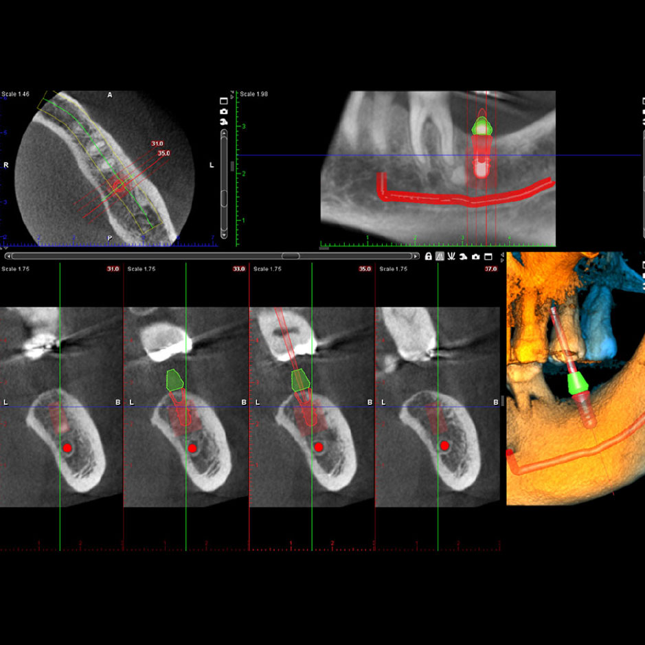
The increasing use of osseo-integrated implants has created a wider demand for radiographic techniques in the pre-surgical and post-operative evaluation of the patient. Correct topographical information is required on the prospective site for planning the implant location.
Our new 3D Dental CT Scan (Cone Beam Technology) goes beyond traditional diagnostics and treatment capabilities by calculating a large volume 3D image set, more than 200 exposures, in a single low-dose 3D scan of 15 seconds or less.
Panoramic radiography for general diagnostics of the tooth arch and the jaw.
Advanced panoramic radiography for specific diagnostics of the tooth arch, the jaw, maxillary sinuses and temporo-mandibular joints.
3D Dental tomographic slices for detailed morphologic diagnostics of facial bones.
Cephalometry for imaging of the skull
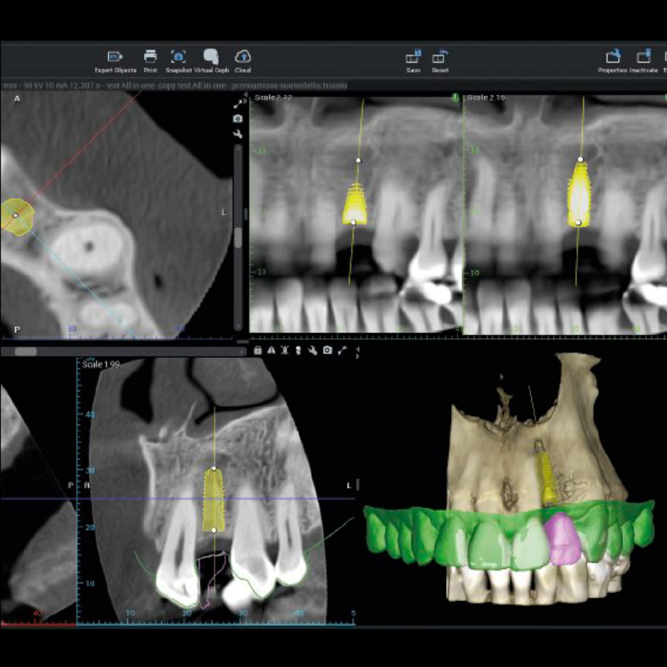
Thanks to 3D pre-operative planning dental implant software we will view axial, sagital and coronal images as well as cross-sections and panoramic images. It allow us to do 3D visualization and simulation of your implant placement and/or bone augmentation procedures before the surgery. With dental implant planning software, surprises during surgery is eliminated and give us an extra margin of safety for our patients.
3D Dental Imaging can be extremely useful in complex cases such as;
Surgical planning for impacted teeth
Diagnosing TMJ or other oral disorders
Dental Implant placement
Reconstructive surgery planning
Evaluation of the jaw, sinus, cavities, nerves and nasal cavity.
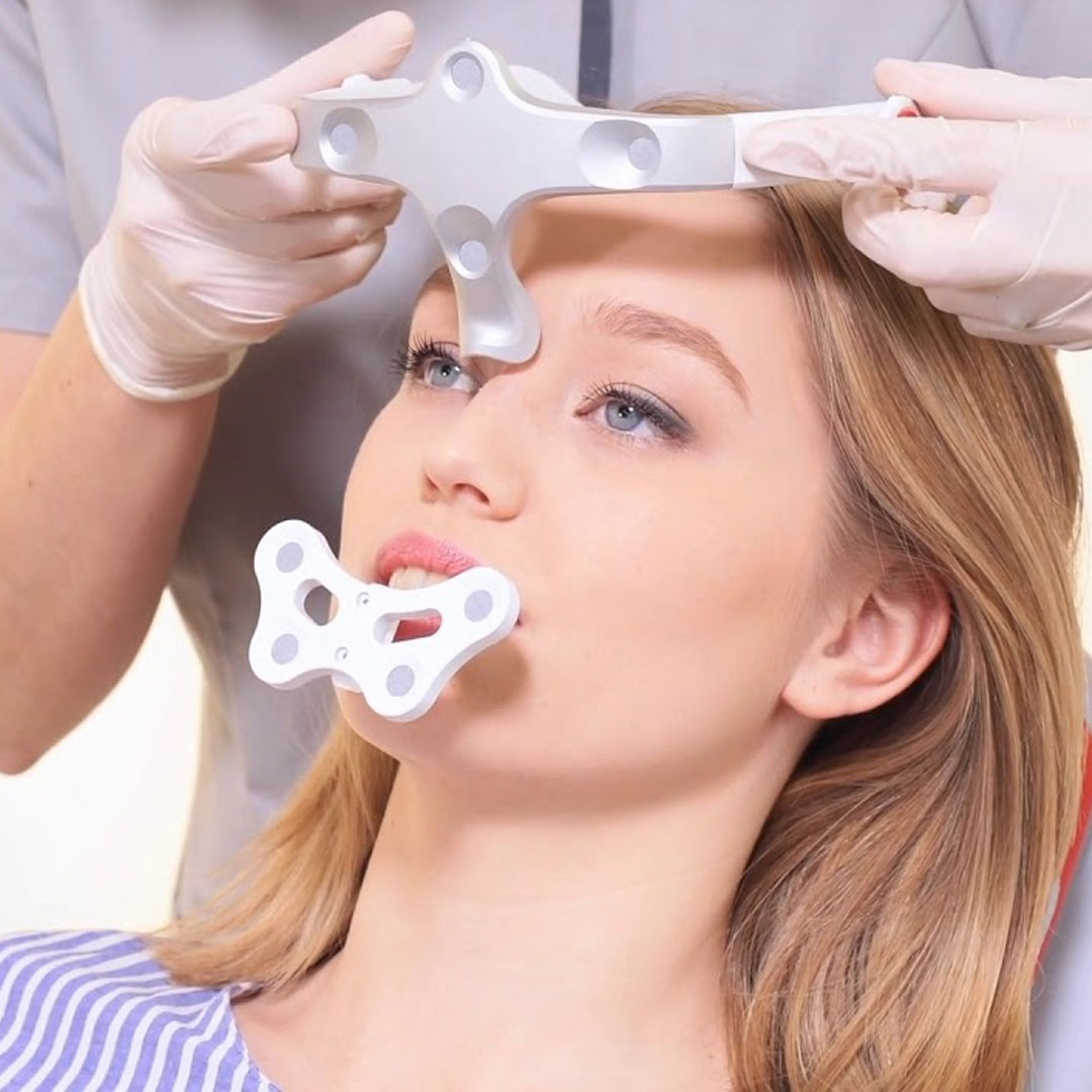
4D DENTISTRY BY MODJAW
NEW DIGITAL DENTISTRY DRIVEN BY 3D MODELING, JAW MOTION AND DYNAMIC OCCLUSION
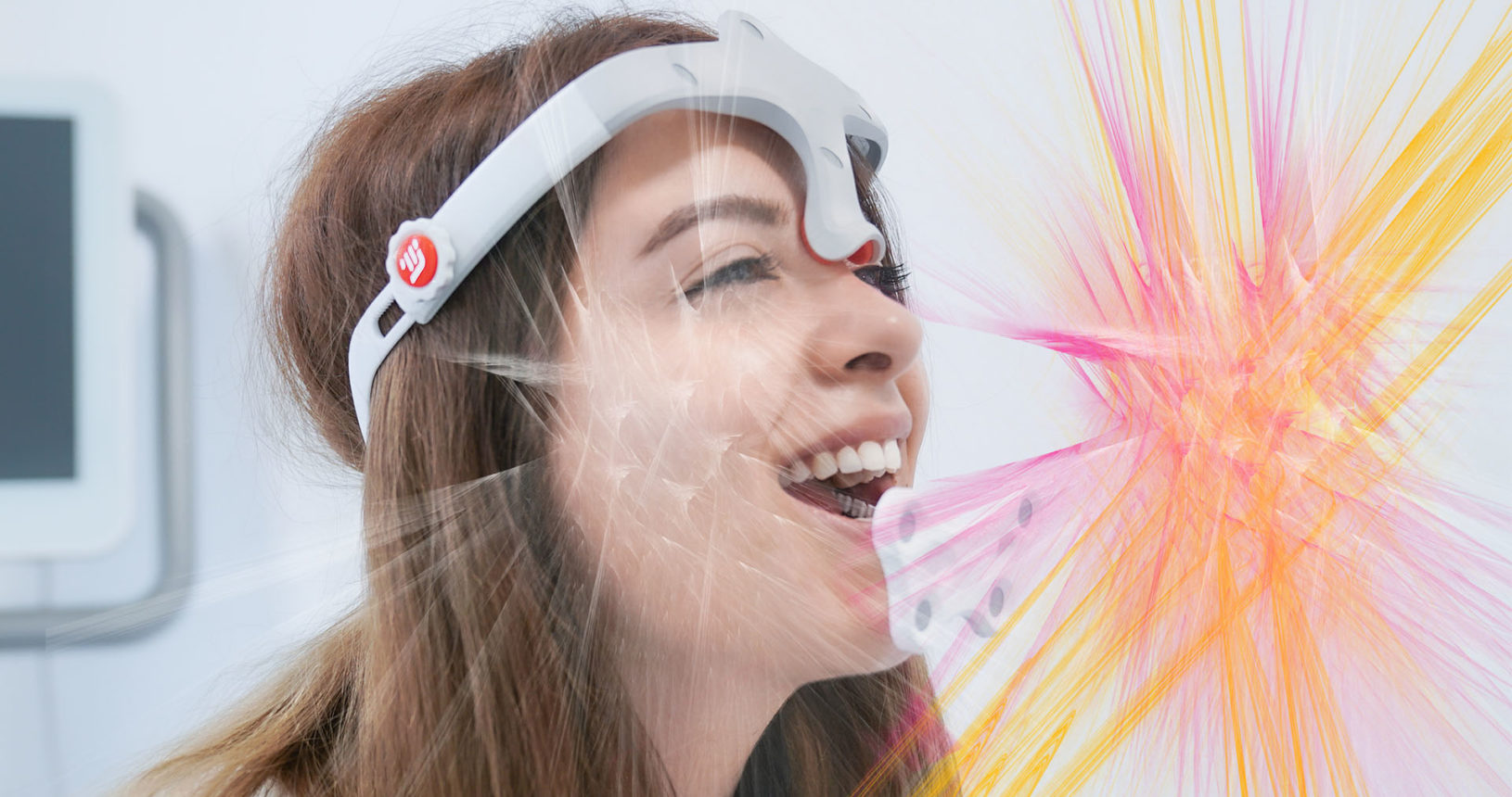
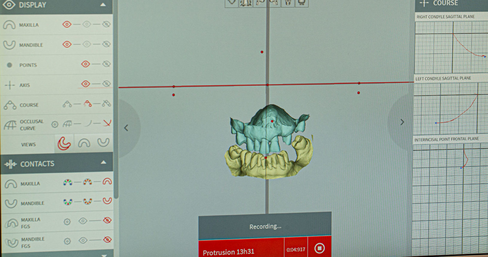
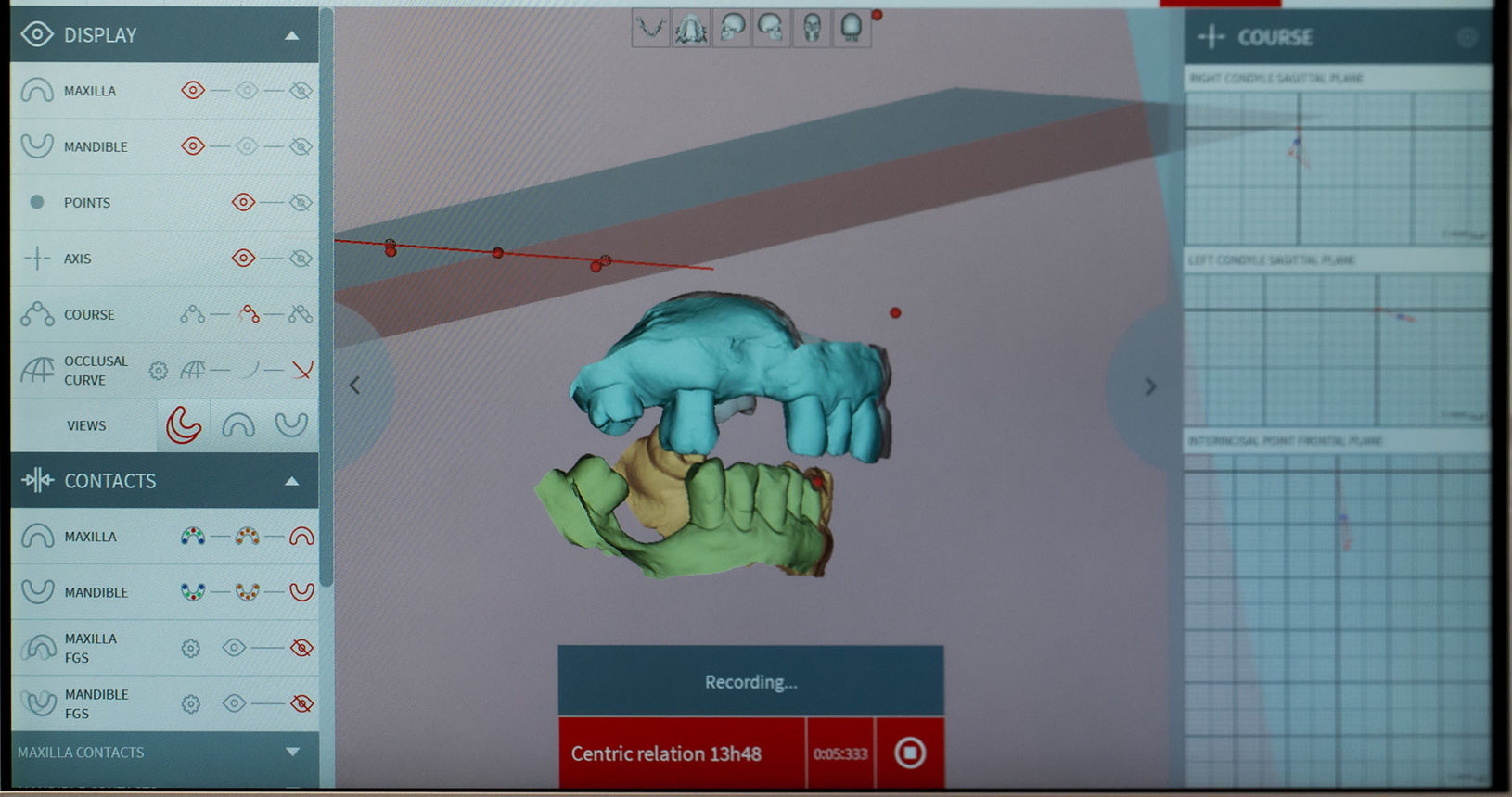
With the 4D dentistry concept, MODJAW introduces a new way of entering your patients’ reality using true jaw motion and dynamic occlusion, in addition to 3D modeling.
4D dentistry is driven by the idea that both static and dynamic parameters should be taken into account to enable complete diagnostics and bespoke restorations.
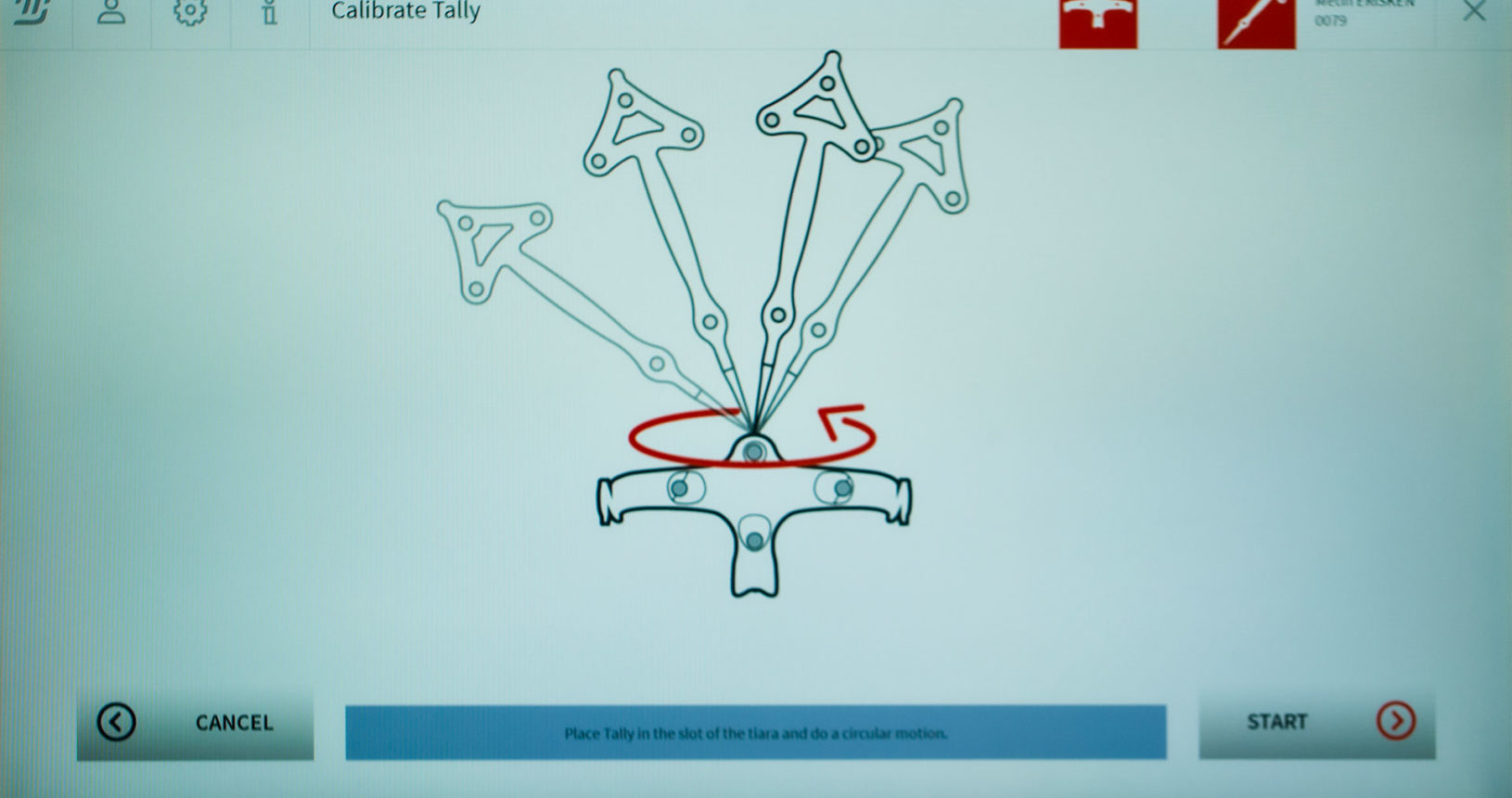
MODJAW’s technology offers a unique experience to you and your patients: efficiency, comfort and advanced interactions. The power of MODJAW Tech in Motion lies in the advanced occlusal analysis : quick, simple and not invasive, with the utmost respect for the health of the patient.
